
Retinal and choroidal imaging refers to advanced diagnostic techniques used to visualize the retina and the choroid (a layer of blood vessels beneath the retina) in great detail. These images are crucial for diagnosing and monitoring various eye conditions, particularly retinal disorders.
Diabetic Retinopathy is a diabetes-related eye condition that affects the blood vessels of the retina, the light-sensitive layer at the back of the eye. It is a leading cause of blindness among adults. The condition occurs when high blood sugar levels damage the blood vessels in the retina, leading to leakage, swelling, or the growth of new, abnormal blood vessels.
Age-Related Macular Degeneration (AMD) is a progressive eye condition that affects the macula, the central part of the retina responsible for sharp, central vision. It is one of the leading causes of vision loss in people over the age of 50. AMD doesn’t cause complete blindness, but it can significantly impair the ability to read, recognize faces, and perform tasks requiring sharp vision.
Retinal Vein Occlusion (RVO) is a condition where one of the veins in the retina becomes blocked, usually by a blood clot. This blockage can cause blood and fluid to leak into the retina, leading to swelling, bleeding, and potential vision loss. There are two main types:
Central Serous Chorioretinopathy (CSCR) is an eye condition where fluid builds up under the retina, specifically in the macula (the central part of the retina responsible for sharp vision). This causes a small detachment of the retina and leads to blurred or distorted central vision.
The analogy of the eye being like a camera helps to understand its function more clearly. The retina, as the “film” of the eye, is essential in capturing the light that enters the eye and translating it into nerve signals for the brain. The optic nerve then acts as the “cable” that transmits these signals to the brain for processing into visual information.
The four key structures — cornea, lens, retina, and optic nerve — are critical for good vision. The cornea and lens focus the incoming light, while the retina captures the image, and the optic nerve transmits it.
Drishti center for advanced eye care Haldwani has a fully-functional retinal department conducting retinal surgeries and medical management of retinal disorders. Managing conditions like diabetic retinopathy, retinal detachment, age-related macular degeneration (ARMD), and trauma is critical, and it’s great to hear that Drishti is equipped to handle these conditions with the latest surgical and medical techniques.
The center’s involvement in pioneering treatments for retinal diseases, particularly advancements for macular degeneration and diabetic retinopathy, shows their leadership in the field.
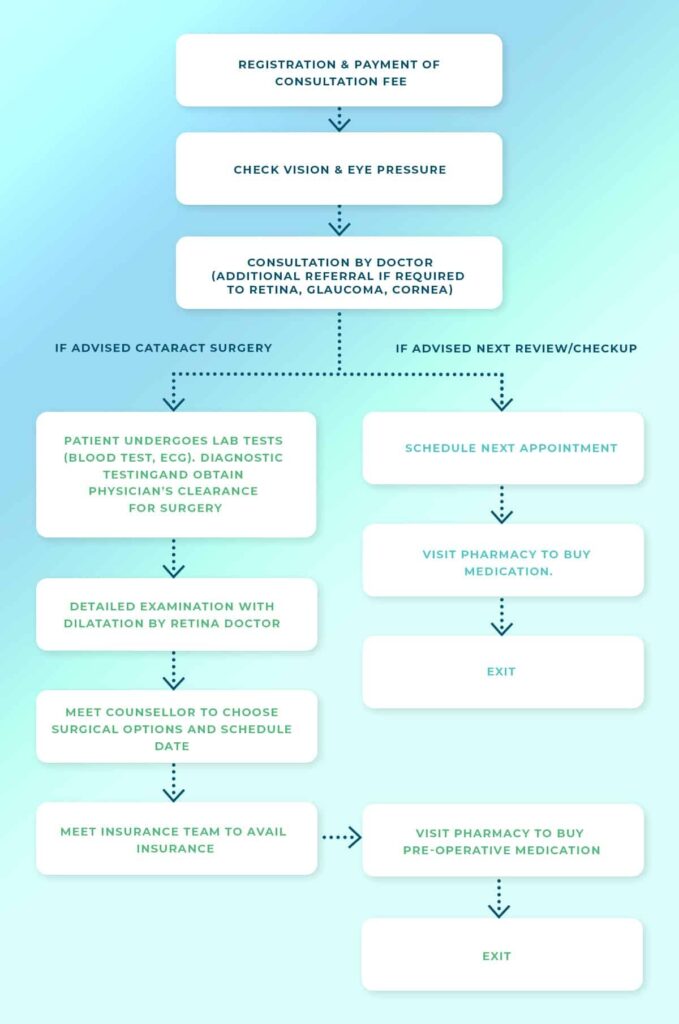
If you have sudden appearance of floaters, blurring of vision, flashes of light in one or both eyes, loss of vision or a “shadow” over the visual field, you should schedule an appointment with an ophthalmologist to get your eyes checked immediately. Your initial consultation with the doctor will approximately take 2 hours if you do not require cross-consultation and up to 4 hours if you require cross-consultation. During your consultation, our doctors and counsellors will determine the best course of action for your visual needs, go over the risks and benefits of treatment, and help you choose the best procedure that is suitable for preserving and improving your vision. We suggest you bring a family member or friend with you to help you with your decision-making.
Retinal surgery refers to surgical procedures used to treat various disorders of the retina, which is the light-sensitive layer at the back of the eye. Retinal surgery is often necessary when conditions threaten vision or cause significant damage to the retina. These surgeries are typically performed by a specialized retina surgeon.
Common Types of Retinal Surgery: