
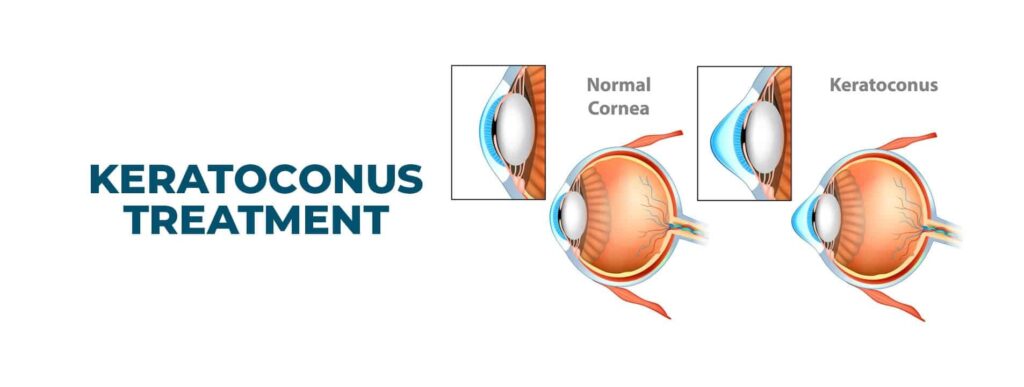
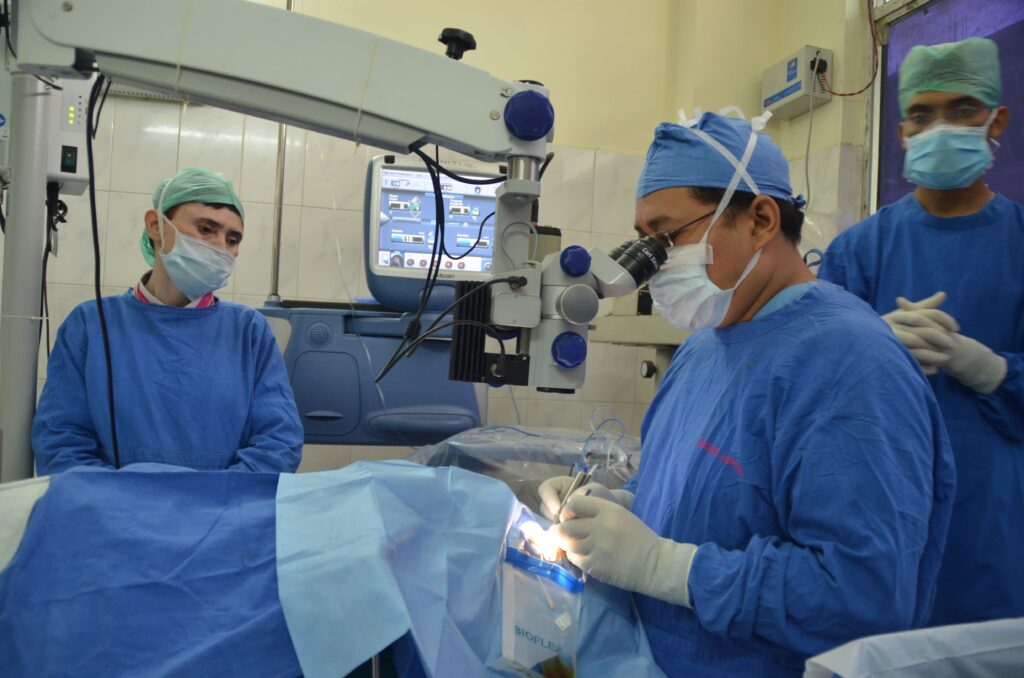
At Drishti Center for Advanced Eye Care, we provide specialized and comprehensive treatment for keratoconus, a progressive eye condition where the cornea thins and bulges into a cone-like shape, leading to distorted vision. As a tertiary care and referral center, we offer the latest advancements in keratoconus management, ensuring optimal outcomes for our patients. Our treatment options include both non-surgical and surgical approaches, tailored to the stage and severity of the condition.
Corneal topography is a procedure used to monitor and measure changes affecting the strength and integrity of the cornea of your eye. At Drishti Center For Advanced Eye Care, Haldwani your cornea will undergo detailed evaluation using all the latest topographers available
Works on the principle of a rotating Scheimpflug camera system for anterior segment analysis. An advanced tool for measuring topography and elevation of the anterior and posterior corneal surface and the corneal thickness – in short the most advanced tool for detection and monitoring progress of your disease.
It consists of a combination of two rotating Scheimpflug cameras and a Placido disk, and allows full analysis of the topography and elevation of the anterior and posterior corneal surface and full corneal thickness. An integrated software also helps us plan your laser surgery based on your corneal map.
Galilei & Orbscan are other topographers used for Keratoconus evaluation.
Advanced software for point to point mapping of your corneal epithelium (first layer of your cornea) helps us in more accurate planning of the laser procedure along with cross linking.
Comprehensive biomechanical screening and Keratoconus detection. Helps determine your eye pressure and the overall strength of the eyeball.
Advanced microscopy technique which enables us to see the ultra microscopic structure of the cornea in just few minutes.


Keratoconus is a condition where the cornea thins and bulges into a cone shape, leading to vision distortion. Its causes are not fully understood, but several factors may contribute:
The condition often progresses gradually, affecting vision over time.
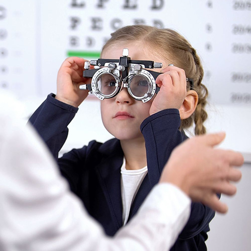
To diagnose Keratoconus, we will review your medical and family history and conduct an eye exam. This also involves other tests to determine more details regarding the shape and strength of your cornea.
We have specially trained optometrists who will use special equipment to check for the right combination of lenses to give you the sharpest vision.
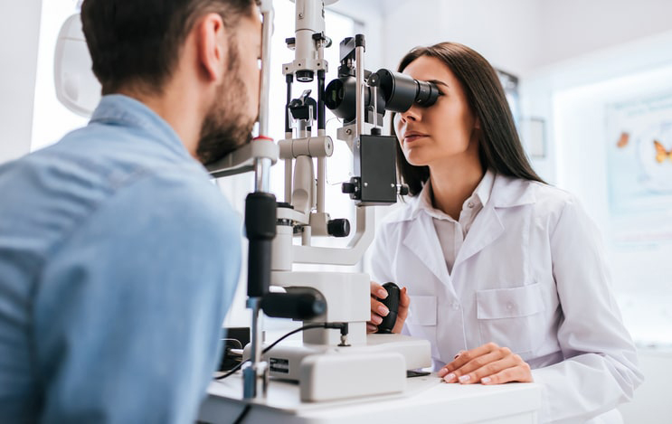
A vertical beam of light is used on the surface of your eye through a low-powered microscope to view your eye. He or she evaluates the shape of your cornea and looks for other potential problems in your eye like allergy etc which have a strong association with keratoconus.
Advanced diagnostic tests, such as optical coherence tomography and corneal topography, record images of your cornea to create a detailed shape map of your cornea’s surface, as well as measure the thickness of your cornea. We also have advanced equipments that help measure the strength of your cornea.
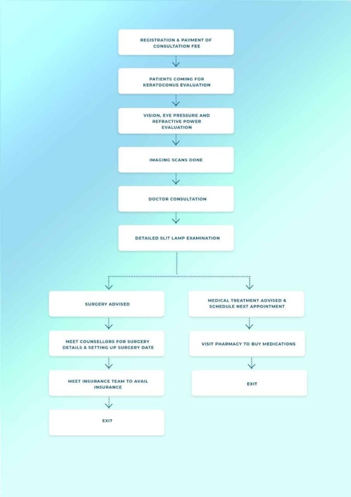
If you have light sensitivity, poor night vision, multiple ghost images, frequent change of glasses or contact lenses, blurring and headaches, you should schedule an appointment with an ophthalmologist to get your eyes checked immediately. Your initial consultation with the doctor will approximately take 2 hours if you do not require cross-consultation and up to 4 hours if you require cross-consultation. During your consultation, our doctors and counsellors will determine the best course of action for your visual needs, go over the risks and benefits of treatment, and help you choose the best procedure that is suitable for preserving and improving your vision. We suggest you bring a family member or friend with you to help you with your decision-making.
Heredity—chances of developing Keratoconus increases if any of your family member is affected.
Overexposure to ultraviolet rays
Poorly fitted contacts
Eye Allergies and chronic eye rubbing-Chronic eye rubbing is associated with developing keratoconus. It may also be a risk factor for disease progression.
Certain diseases, including retinitis pigmentosa, atopic disease and connective tissue disorders.
Down Syndrome
Lorem ipsum dolor sit amet, consectetur adipiscing elit. Sed auctor turpis eu arcu sagittis, id sagittis justo suscipit.